
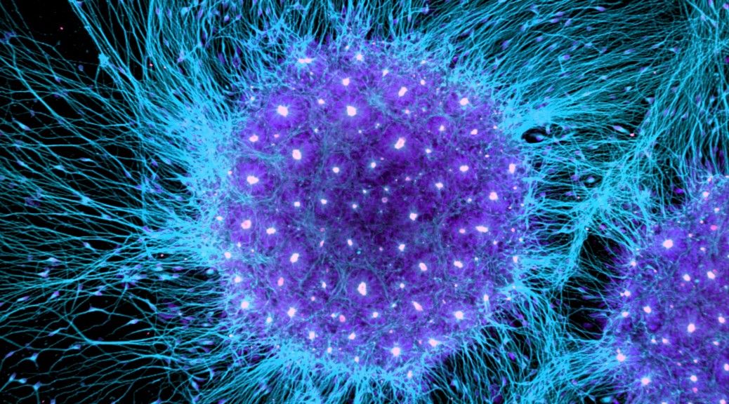
Looking through the lens of a microscope will give you a glimpse of a new and practically invisible world. And it can be breathtaking. The Nikon Small World contest celebrates the most stunning microscope photos from scientists around the world and 2016′s competition had some spectacular entries. From the foot of a diving beetle and dried coffee crystals to brain cells and (believe it or not!) cow dung, here are this year’s winning images from an unseen universe: Four-day-old zebrafish embryo View photos 1st place (Dr Oscar Ruiz/Nikon Small World) More Polished slab of a Teepee Canyon agate View photos 2nd place (Douglas L Moore/Nikon Small World) More Brain cells from skin cells View photos 3rd place (Rebecca Nutbrown/Nikon Small World) More Butterfly proboscis View photos 4th place (Jochen Schroeder/Nikon Small World) More Read More Front foot of a male diving beetle View photos 5th place (Dr Igor Siwanowicz/Nikon Small World) More Air bubbles formed from melted ascorbic acid (vitamin C) crystals View photos 6th place (Marek Mis/Nikon Small World) More Leaves of Selaginella (lesser club moss) View photos 7th place (Dr David Maitland/Nikon Small World) More Wildflower stamens View photos 8th place (Samuel Silberman/Nikon Small World) More Espresso coffee crystals View photos 9th place (Vin Kitayama & Sanae Kitayama/Nikon Small World) More Frontonia (unicellular protozoan) View photos 10th place (Rogelio Moreno Gill/Nikon Small World) More Scales on the underside of a butterfly wing View photos 11th place (Francis Sneyers/Nikon Small World) More Human hela cell (immortal cells used in scientific research) undergoing cell division View photos 12th place (Dr Dylan Burnette/Nikon Small World) More Poison fangs of a centipede View photos 13th place (Walter Piorkowski/Nikon Small World) More Retinal ganglion cells of a mouse View photos 14th place (Dr Keunyoung Kim/Nikon Small World) More Head section of an orange ladybird View photos 15th place (Geir Drange/Nikon Small World) More Radiolarians (zooplankton) carefully arranged by hand View photos 16th place (Stefano Barone/Nikon Small World) More Slime mould View photos 17th place (Jose R Almodovar/Nikon Small World) More Abdominal segment and hind leg of a broad-shouldered leaf beetle View photos 18th place (Pia Scanlon/Nikon Small World) More Human neural rosette primordial brain cells, differentiated from embryonic stem cells in the culture dish View photos 19th place (Dr Gist F Croft, Lauren Pietilla, Stephanie Tse, Dr Szilvia Galgoczi, Maria Fenner and Dr Ali H Brivanlou/Nikon Small World) More Cow dung View photos 20th place (Michael Crutchley/Nikon Small World) More For more images check out Nikon Instruments’s Instagram account .
From : uk.news.yahoo.com